
What is known about the protein structure and
function?
| The hMLH1 protein has 756 amino acids and has been completely sequenced. The protein has 2 conserved regions at the amino terminus. In other words, there are two sections of the protein that are nearly identical in the proteins of other organisms. (Ref. 2) These amino acid sequences are (with single letter abbreviations) KELVEN and GFRGEA. The hMLH1 protein is 41% identical to the yeast DNA mismatch gene yMLH1, with the last 13 amino acids being identical between the two proteins. The protein is composed of 19 exons, 50% of all MLH1 mutations that have been described occur in exons 15 and 16 (Ref. 2) The first seven exons are highly conserved between MLH1 and PMS1. | 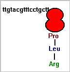 |
One proposed explanation for the increased frequency of mutation is dinucleotide repeat instability. In areas of DNA where two nucleotides are repeated, there is an increase in spontaneous mutation within these repeats. (Ref. 2) This can lead to more than a 100 fold increase in the mutation rate (Ref. 8). Mutations resulting from dinucleotide repeat instability are common in HNPCC families. Consistent with this idea is the evidence from a study showing that the HNPCC tumor DNA had different markers than the normal tissue DNA indicating replication error. (Ref. 1)
An issue that is still under considerable investigation
is why the colon is the most frequent target for tumor development. Mismatch
repair genes are housekeeping genes, meaning they are expressed in nearly
every cell and are essential for normal cell function. It is not yet understood
why tumor development is restricted to certain organs. One possibility
is that the colon mucosal layer has more exposure for long time periods
to carcinogens found in food, toxic metabolites, and bacterial products.
Another possibility is that the colon mucosal layer has a high proliferation
rate which could overburden and exhaust the mismatch repair genes. This
might possibility would also hold true for the endometrial mucosal
layer.
 |
Unlike diseases like Down Syndrome, HNPCC is not caused by a specifically recognized mutation. There are 4 genes that have been linked to HNPCC, and for each of those genes there is a multitude of mutations that can occur within the genetic code. This graph shows the extent of the heterogeneity of HNPCC. In other words, the slice of the graph is proportional to the number of different of genetic mutations that give rise to the same disease. HNPCC ranks second in hereditary colon cancers. |
A study completed in 1996 using mice showed that
the MLH1 protein was not only involved in DNA mismatch repair, but also
in meiotic crossing over. Mice that were created with a mutation in the
MLH1 gene not only showed mutations that are associated with HNPCC, but
resulted in sterility of both males and females. Sterility is usually associated
with disruption in the normal meiotic process. They found evidence showing
MLH1 located at chiasmata sites on meiotic chromosomes. Chiasmata are the
cross-shaped structures seen during crossing over of chromosomes in meiosis.
No other studies have been found that support this result, and sterility
has not been associated with HNPCC in humans. (Ref.
2)
What other sources about the protein are available?
|