|
Links |
Linkage Studies | |
|
Initial Discovery and Clinical Aspects Molecular/Biochemical Properties and Characteristics of Cri Du Chat-Related Proteins Links to Cri Du Chat Support Groups
|
Since the discovery of the genetic disease Cri du Chat, research around the world has been dedicated to linking certain genes to the causation of specific phenotypes common to this syndrome. One of the first studies, however, was preformed Jérpome Lejeune, whose work was dedicated to finding the chromosome responsible for the genetic disease. Below is an excerpt from his paper “Mental and Physical Deficiency related to a partial deletion of the short arm of chromosome 5.” Here, Lejeune had studied the karyotypes of different individuals showing certain symptoms and characteristics of CdCS, trying to determine what chromosome, if any, are responsible for this disease: “The first patient, studied around a year ago, was referred to us by Dr. Vialatte. This poorly developing girl, severely retarded, had a few malformations, but as reported by the pediatrician, she had a very peculiar cry, quite similar to the mew of a cat. Physically, this girl was under developed (Birth weight 1.700 g height (41 cm), micro cephalic (29.5 cm of cranial perimeter) and had a broken round face. The only malformation noted were, marked hypertelorism, epicanthus, slanting eyes, small mandibles and low set ears, otherwise normal. The cry was indeed very peculiar: a rather high pitched prolonged and weak sound was emitted by the girl when disturbed. Its plaintive tonality was strikingly similar to the mew of a suffering cat. The skin biopsy showed 46 chromosomes apparently normal, but one of the members of the group 4 to 5 was exhibiting a short arm much too short. All the 40 cells examined showed this characteristic element (see fig. 1 and 2).
The second case was recognized and diagnosed clinically by Professor La Fourcade. The physical picture was identical: a broad, round face, a flat bridge of the nose, hypertelorism, epicanthus; slanting eyes, small mandibles and low set ears. The "cri du chat" was obvious. The chromosomes of this boy showed the same abnormality, a loss of around one half of the short arm of chromosome 5.
The third patient, a girl, was detected by Dr. Berger the same week and the characteristic abnormalities were recognized. Microcephaly and round face, hypertelorism, epicanthus, slanting eyes, low set ears, and small mandibles. The chromosomes were identical. One of the members of group 4 to 5 had very reduced small arm.
Thus using deliberately and purposely a tautological
approach, we would say the disease is due to the loss of around one half
of the short arm of chromosome 5 and, conversely, we would state, by
definition, that we call 5 the chromosome in which the loss of part of the
short arm produce the "cri du chat" syndrome. It is very likely many of
those genetic changes are not directly related to the cerebral machinery
but are just interfering with its functioning.” Another researcher, J. Overhauser (1994) analyzed the 5p deletion breakpoints in 49 individuals using somatic cell hybrids. His group used 5p-specific DNA probes to order most of the chromosomal breakpoints present by hybridization to somatic cell hybrid DNA. Comparisons between the deletions present in the patients and their clinical features identified several chromosomal regions that were involved in specific clinical features. A critical chromosomal region involved in the high-pitched cry mapped to proximal 5p15.3, while the chromosomal region involved in the remaining features of the syndrome mapped to a small region within central 5p15.2. This latter region was estimated to be about 2 Mb in size. Deletions that did not include these 2 chromosomal regions presented varying clinical phenotypes from severe mental retardation and the characteristic small head to a clinically normal phenotype. (OMIM)
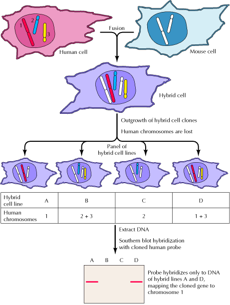
Figure 4.27. Somatic cell hybridization Human and mouse cells in culture are fused by treatment with a virus or chemical agent, yielding a hybrid cell containing both human and mouse chromosomes. (In this example only three human and two mouse chromosomes are shown.) Human chromosomes are unstable in such hybrids and are gradually lost during the outgrowth of individual clones of the hybrid cells. Therefore, a panel of hybrid cell lines is obtained, with each line containing a different complement of human chromosomes. Hybridization of a cloned gene to DNAs extracted from such a panel of hybrid cell lines can be used to map the gene to a specific human chromosome. In this illustration, a human probe hybridizes to DNAs of cell lines A and D, but not B and C. Since only hybrid cell lines A and D have retained chromosome 1, these results map the cloned gene to this human chromosome. picture and caption taken from Geoffrey M. Cooper at http://www.ncbi.nlm.nih.gov/books/bv.fcgi?rid=cooper.figgrp.650 |